

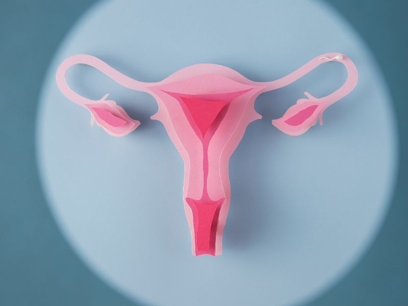
The uterus, one of the main organs of the female reproductive system, is held in a fixed position in the body by certain connective tissues and the pelvic floor muscles. Pelvic support tissues that weaken over time, childbirth, and hormonal changes can make it harder for the uterus to stay in place. In this case, the uterus drops downward, and this condition is called uterine prolapse.
In its mild form, this condition usually causes no symptoms, but as it progresses it can seriously affect quality of life, both physically and psychologically. Uterine prolapse is seen not only at an older age but also in young women who have given birth. That is why it is extremely important to recognize the symptoms, get the right diagnosis, and start treatment on time.
What Is Uterine Prolapse?
Uterine prolapse is medically called "uterine prolapse (prolapsus uteri)" and is a condition in which the uterus drops down from its normal position into the vaginal canal. In women of reproductive age, the uterus is about 5-7 cm long and sits inside the pelvis, behind the bladder and in front of the rectum. Normally it is held in place by strong pelvic muscles and connective tissues. However, weakening of these support systems can cause the uterus to move downward.
What Causes Uterine Prolapse?
The main cause of uterine prolapse is that the muscles and connective tissues in the pelvic region weaken over time and can no longer adequately support the uterus. This weakening usually occurs as a natural result of aging. However, it is not limited to age alone; various physical and hormonal factors can also set the stage for uterine prolapse.
In particular, having many vaginal births puts repeated pressure on the muscles and connective tissues of the pelvic floor, causing these structures to weaken. Similarly, giving birth to a large baby or having a difficult delivery can also disrupt the structural integrity of the tissues. During menopause, the drop in estrogen levels causes the muscles and connective tissues to lose their elasticity, which increases the risk of prolapse.
Conditions that put constant pressure on the pelvic floor, such as chronic constipation and coughing, can also make prolapse more likely. Excess weight and obesity can raise intra-abdominal pressure and lead to downward displacement of the uterus. Previous surgery on the pelvic region, pelvic organ tumors, genetic connective tissue weakness, and smoking are among the other factors that increase the risk of uterine prolapse.
How Can You Tell If You Have Uterine Prolapse?
Uterine prolapse may not cause the same symptoms in every woman. Mild prolapse in particular usually does not cause noticeable complaints and is often discovered by chance during a routine gynecological exam. However, in advanced cases where the uterus has dropped further, some physical symptoms that affect daily life may appear.
Women usually complain of a feeling of internal pressure or heaviness in the vaginal area. In some cases, there is a feeling as if tissue is coming out of the vagina, and this may also become physically visible. Straining to urinate, frequent urination, urinary incontinence, or a feeling that the bladder has not emptied completely are also common signs of uterine prolapse. In addition, pain during sexual intercourse, a feeling of pressure in the vaginal area, increased vaginal discharge, or spotting-type bleeding may also occur. Some patients also complain of difficulty with bowel movements, constipation, or a feeling of pressure in the rectum.
Symptoms of Uterine Prolapse
Uterine prolapse can cause symptoms of varying severity depending on the degree of prolapse. While mild cases may cause no symptoms, moderate and advanced prolapse can cause physical complaints that lower quality of life. The most common symptoms of uterine prolapse are:
- A mass or tissue coming out of the vagina: Patients usually describe a “bulge” or a feeling of fullness at the vaginal opening.
- A feeling of pressure or heaviness in the vagina: A feeling of pelvic pressure is seen that increases especially after standing for a long time or at the end of the day.
- Urinary incontinence or straining to urinate: Because of the pressure on the bladder, urine flow may become difficult or involuntary urine leakage may occur.
- A feeling of incomplete bladder emptying: Some patients report a feeling of fullness in the bladder after urinating.
- Frequent urination or waking up often at night to urinate (nocturia): Pressure on the urinary tract can create the need to go to the bathroom often.
- Constipation or straining during bowel movements: Pressure on the rectum can disrupt bowel movements.
- Pain or discomfort in the lower back or groin: Pain that increases after standing for a long time or after physical activity may occur.
- Pain during sexual intercourse (dyspareunia): Because the position of the uterus is disrupted, sexual intercourse can be uncomfortable.
- Increased vaginal discharge or bleeding between periods: Friction or injury to the vaginal tissues can cause discharge and bleeding.
- Hygiene problems and increased risk of infection: Irritation of the protruding tissues can set the stage for yeast or bacterial infections.
How Is Uterine Prolapse Diagnosed?
Uterine prolapse is diagnosed with a pelvic exam performed by a specialist gynecologist. During this exam, the patient is usually encouraged to bear down, cough, or squeeze her pelvic muscles as if holding in urine, so that the prolapse can be evaluated more clearly. These maneuvers help show how far the uterus has descended into or out of the vagina. The degree of prolapse can be observed through the physician's manual evaluation during the exam.
Uterine prolapse is usually classified into four stages:
- Grade 1: The uterus has descended slightly and moves down into the vagina.
- Grade 2: The uterus has reached the vaginal opening.
- Grade 3: Part of the uterus has come out of the vagina.
- Grade 4 (Total Prolapse): The uterus has come out completely.
In some cases, not only the uterus but also other pelvic organs such as the bladder (cystocele) or rectum (rectocele) may prolapse along with it. To clarify such combined conditions and create a complete anatomical map of the prolapse, advanced imaging techniques such as transvaginal ultrasound, pelvic MRI, or dynamic magnetic resonance imaging are used. These tests are especially important for providing detailed information in patients for whom surgery is being planned.
How Is Uterine Prolapse Treated?
The treatment of uterine prolapse is planned individually according to the severity of the prolapse, the patient's age, overall health, and its impact on quality of life. For mild uterine prolapse, conservative (nonsurgical) methods are usually preferred without the need for surgery. At this stage, Kegel exercises to strengthen the pelvic floor muscles are recommended. At the same time, lifestyle changes such as losing excess weight and getting chronic constipation and coughing under control are also an important part of treatment.
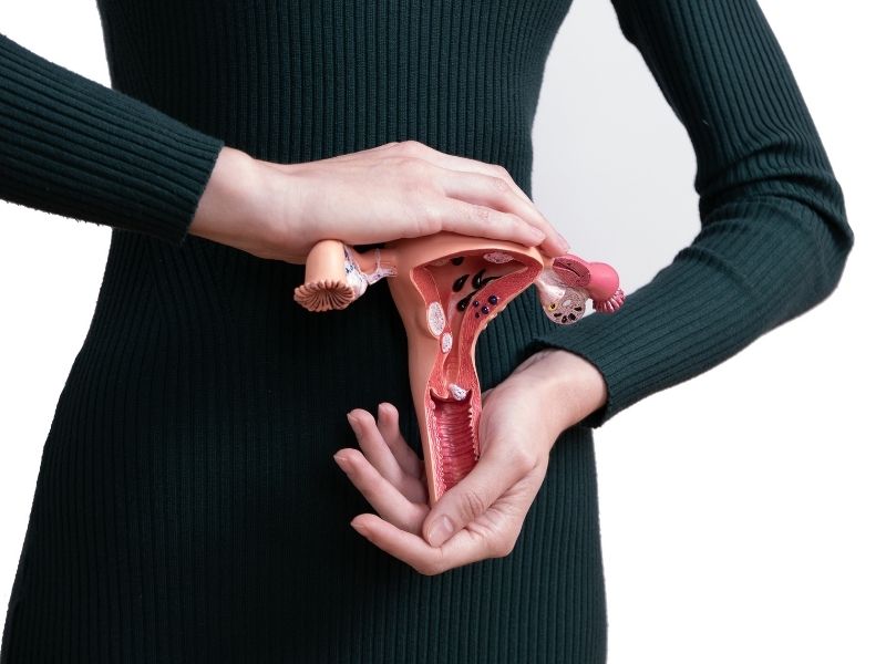
For moderate and advanced prolapse, more effective treatment methods may be needed. As a nonsurgical option, a pessary, a silicone ring placed inside the vagina, can be used. This device supports the uterus and can keep the prolapse from progressing, and it is a suitable solution especially for patients who do not want surgery or who are at risk from surgery.
However, in severe cases, surgery is the most effective solution. With this surgery, the weakened pelvic support tissues are repaired and the normal anatomical position of the organs is restored. In some advanced cases, a hysterectomy, meaning complete removal of the uterus, may be performed. The surgical method is carefully planned, taking into account the patient's age, fertility plans, and overall health.
Uterine Prolapse Surgery
Uterine prolapse surgery is performed to repair the weak pelvic support tissues that cause the uterus to drop toward the vagina. Depending on the degree of prolapse, the options of keeping the uterus in place (suspension) or removing the uterus completely (hysterectomy) are considered.
The surgery can be performed with three different techniques: vaginally, laparoscopically (minimally invasive), or as open abdominal surgery. Vaginal hysterectomy is the most commonly preferred method; the uterus is removed through the vagina and the support structures are repaired. In the laparoscopic technique, special instruments are used through small incisions made in the abdominal wall, and the patient recovers faster.
In some cases, with supportive procedures such as sacrocolpopexy, the vaginal vault is suspended with a synthetic mesh to strong connective tissue on the spine. The surgery usually takes 1-2 hours, the hospital stay is 1-3 days, and recovery takes about 4-6 weeks. For detailed information and support, you can consult Ataşehir Obstetrician-Gynecologist Prof. Dr. Çiğdem Yayla Abide.